
From Dr David Gunn’s teaching slides
What is the perfect first case?
A clear visual goal. A decentered cone. One segment over the steepest area.
One eye sees reasonably well
The fellow eye provides useful vision, while the poorer eye has a clear visual problem.
A poorer eye worth improving
The slide’s starting profile is CDVA < 6/12–6/18 in the poorer eye.
Contact lenses are not working
Contact-lens intolerance gives the patient a practical reason to seek better vision.
Modest expectations
Aim for useful improvement. The patient understands that glasses or contact lenses may still be needed.
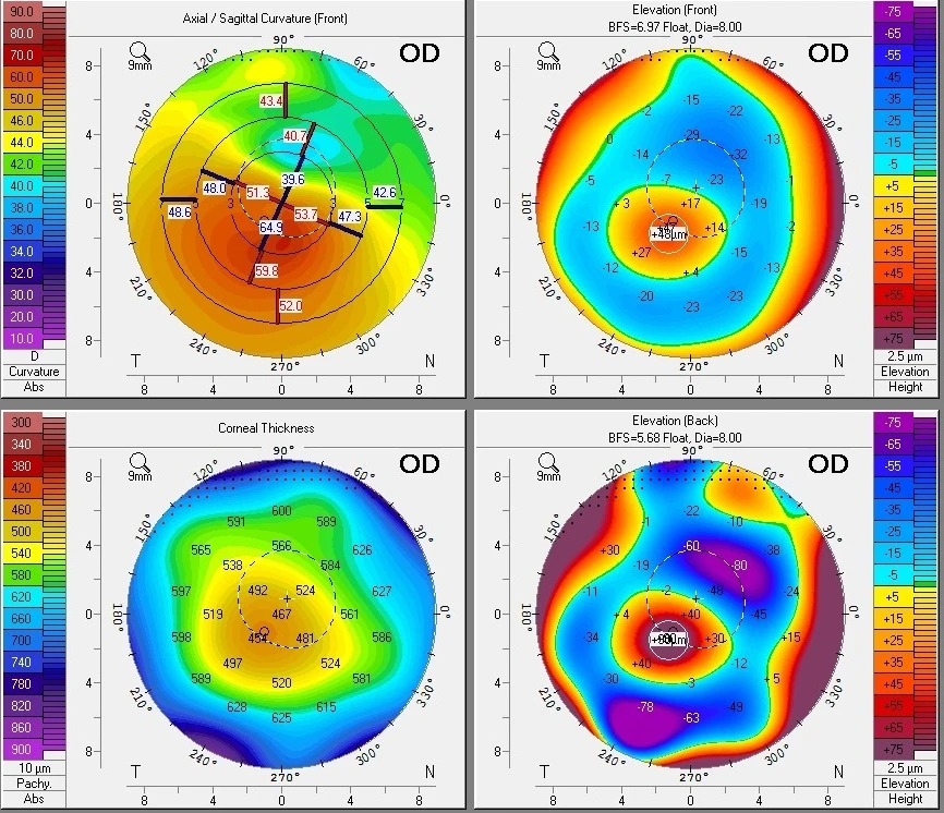
The pattern
A decentered cone
Find the steep region on the curvature map, then read it alongside pachymetry and elevation.
The starting plan
One segment over the steepest area
A straightforward single-segment plan keeps the first case easier to understand and reproduce.
This is a first-case teaching profile. Final selection still depends on the whole clinical assessment and a plan reviewed with an experienced CAIRS colleague.
Start with the patient’s visual problem
The useful question is what the patient needs to improve. Poor quality vision in spectacles, irregular astigmatism and difficulty wearing rigid or scleral contact lenses are common reasons to consider CAIRS. Record refraction, uncorrected and corrected acuity, lens tolerance and the symptoms that matter to the patient.
A patient may read a reasonable line on the chart and still describe ghosting or poor visual quality. Equally, a striking topography does not automatically make a good first case. Discuss the intended improvement, the possibility of continued glasses or contact lenses, and the potential need for further treatment.
Choose a map you can explain
Start with reproducible tomography and a recognisable cone pattern. Review curvature and pachymetry together, and use an absolute colour scale when comparing severity between eyes. An automatically rescaled map can make a mild and a severe cone look deceptively similar.
Ask three questions from the first-case teaching:
- Where is the cornea steep? Locate the steep region and its extent rather than relying on Kmax alone.
- Does the pattern suggest one or two segments? An inferior or decentered cone and a more central, symmetric pattern may need different distributions of tissue.
- How much tissue is appropriate? Use a nomogram that matches the preparation method and dimensions of the implant you intend to use.
For an early case, choose a plan that is straightforward to describe and reproduce. Markedly asymmetric cones, complex tapers and unequal paired segments add decisions that become easier with experience.
Leave the difficult cases until you have experience
Dr Gunn’s teaching highlights post-LASIK ectasia, very steep central corneas, very thin corneas and central apical scars as cases to avoid initially. These eyes may warrant CAIRS assessment by an experienced surgeon, but they add complexity to selection, channel placement and expectations.
That teaching slide uses central K above 60 D and CCT below 330 µm as beginner caution flags. These are first-case teaching points, rather than universal eligibility limits; central keratometry, Kmax, CCT and thickness along the planned channel are different measurements.
Inspect the ocular surface and address active surface disease, allergy and eye rubbing. Assess the cornea’s thickness along the whole proposed channel, alongside central and thinnest-point measurements.
Make the progression plan explicit
CAIRS aims to improve corneal shape and visual function. Review progression and decide separately whether and when cross-linking is needed. Document prior CXL because it can influence the cornea’s response, and discuss sequencing with the colleague helping you plan the case.
Before you book your first case
- Have an experienced CAIRS surgeon review the maps and proposed plan.
- Confirm the tissue source, preparation method, dimensions and availability.
- Check the channel and implant parameters together in CAIRSPlan.
- Practise tissue handling and insertion with your intended instruments in a wet lab.
- Agree the visual goal and arrange postoperative review, with a pathway for adjustment or complications.
A good first case has a clear reason for surgery, a plan you understand and practical support in theatre.
Teaching and further reading
Drawn from Dr David Gunn’s “What is the perfect first case?” and “Starting CAIRS — Planning your first case” slides, together with the CAIRSPlan teaching library.