
If you are making the channels manually, you will need to buy the appropriate dissectors for your planned channel geometry. Add separators, pullers or forceps to suit your insertion and handling technique.
Tools for making the donor implant are a separate choice: manual trephination and customisation need their own preparation instruments.
Cronin–Gunn instruments
Developed with Dr Brendan Cronin and Dr David Gunn. The titanium set follows a 6 mm channel curve, with clockwise and counter-clockwise configurations.
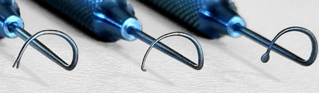
Cronin–Gunn CAIRS set
Show full instrument photo
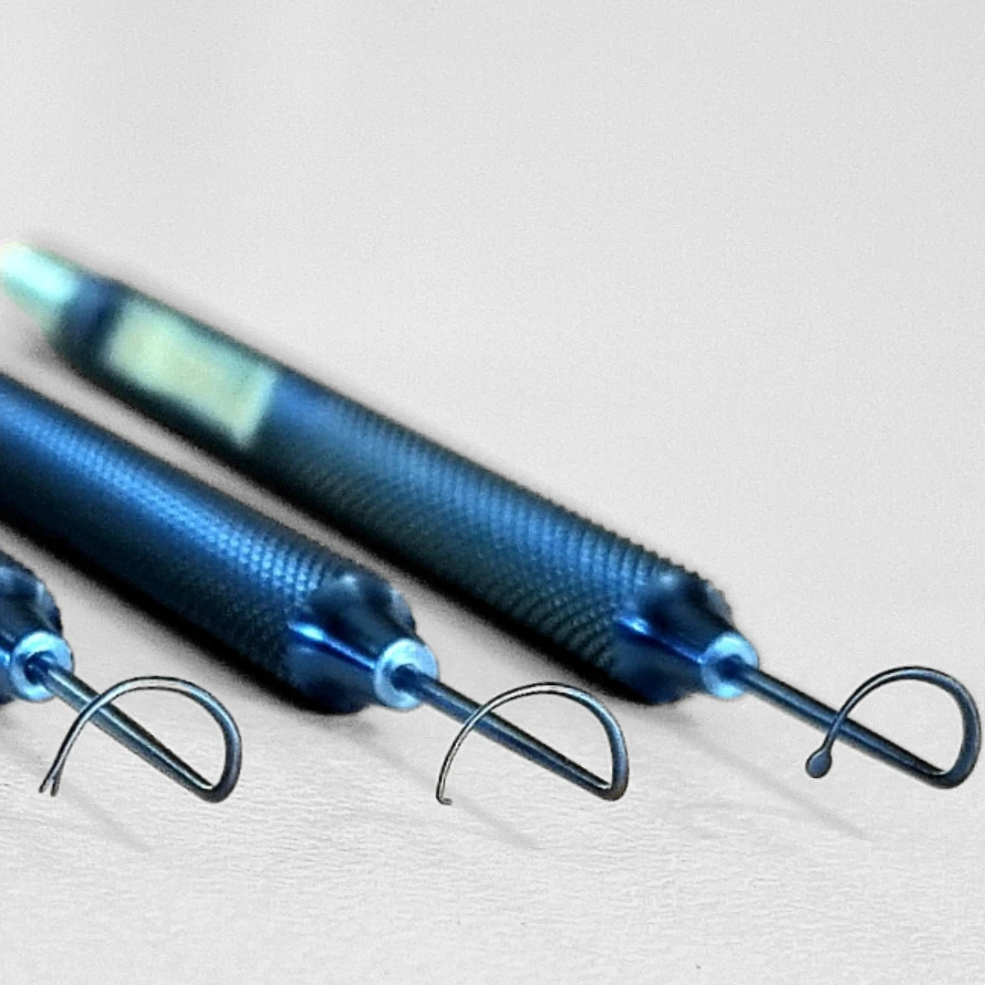
SRB Devices
Separator, pusher and puller for opening the channel, advancing the segment and drawing it into position.
Choose the directional instruments for the insertion path you have planned.
Dr Gunn and Dr Cronin have no financial interest in these instruments.
Soosan Jacob’s trephines and customiser
The trephine creates the donor ring. The rounded customiser helps transfer a personalised shape and arc-length plan onto the tissue.
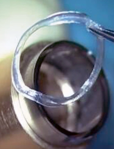
Jacob CAIRS trephines
Show full instrument photo
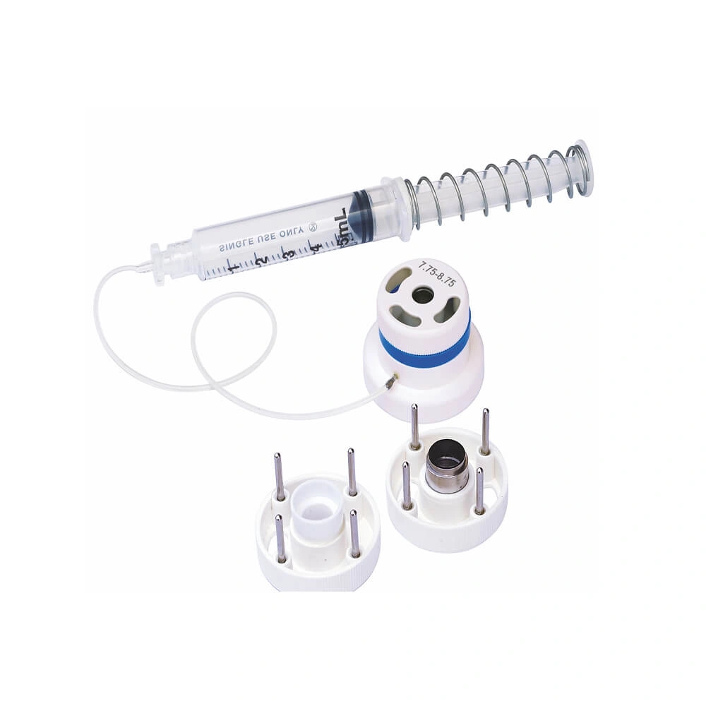
Madhu Instruments
Two concentric blades cut a donor stromal ring for manual preparation. The blade pair determines the annular width before the strip is orientated and customised.
The 2025 catalogue lists inner / outer diameter pairs of 5.50 / 9.50, 6.50 / 8.00, 7.75 / 8.75 and 8.00 / 9.50 mm. Check the current catalogue for the required size.
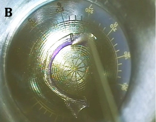
Jacob CAIRS customiser
Epsilon USA · EB-39
The round, domed template — the “ball” — is used to mark arc length and taper transition points before manually shaping the segment.
Align the tissue to the planned optical zone and meridians, mark the intended shape, then trim on a suitable cutting surface using the trained technique.
Photograph: Soosan Jacob.
Awwad CAIRS instruments · Epsilon
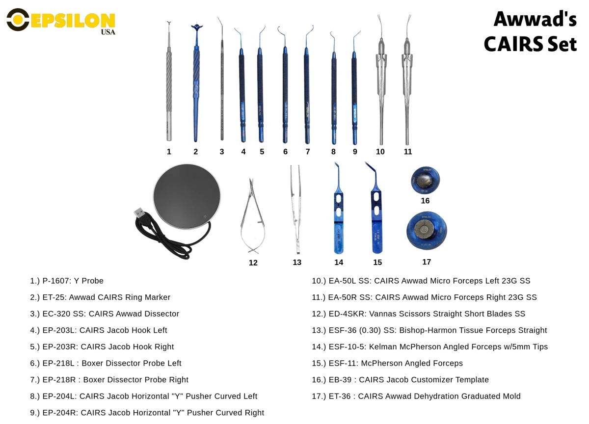
Epsilon’s set brings together marking, dissection, preparation and insertion tools, including the Awwad micro forceps and graduated dehydration mould.
Awwad CAIRS micro forceps
Show full instrument photo
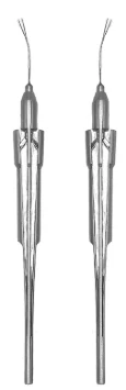
Epsilon USA · EA-50L SS / EA-50R SS
Left and right 23G stainless-steel micrograspers for controlling and adjusting the allogenic segment within the tunnel.
Dr Awwad’s short video demonstrates these dedicated CAIRS forceps.
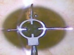
Awwad CLEAR marker
Show full instrument photo
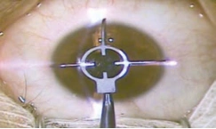
Epsilon USA · ET-32
The three-blade marker for CLEAR transfers centration and axis references onto the cornea before femtosecond laser docking.
Centre the marking reference on the intended light reflex and maintain that reference through docking. The photo shows the marker in use.
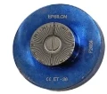
Awwad dehydration mould
Epsilon USA · ET-36
A graduated circular mould that supports the segment’s arc shape during dehydration and provides degree references for preparation.
Choose the geometry and hydration workflow that match the intended implant and channel.
Also listed in the set: a Y probe, CAIRS dissector, left/right Jacob hooks and Y pushers, Boxer dissector probes, scissors and tissue forceps. See the complete Epsilon set →
Aylin Kılıç · AK instruments
AK instruments were developed by Dr Aylin Kılıç for the KeraNatural CAIRS workflow. The instrument guide lists Optimal Sağlık, Istanbul, as the distributor.
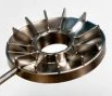
AK Marker
Show full instrument photo
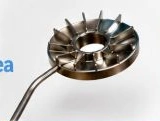
Aylin Kılıç · Optimal Sağlık
A marker for transferring the planned start, end and arc length onto the cornea, using 30-degree sectors to reproduce the topographic orientation.
Photograph from Dr Kılıç’s own introduction of the instrument. Ask the distributor for the current specifications.

AK forceps
Show full instrument photo
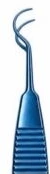
Aylin Kılıç · Optimal Sağlık
Titanium forceps for tissue handling and implantation in the KeraNatural workflow. The curved working ends help control the tissue during insertion.
Photograph from Dr Kılıç’s own introduction of the CAIRS set.
AK tunnel separators
Show full instrument photo
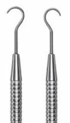
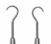
Aylin Kılıç · Optimal Sağlık
Right and left stainless-steel separators for accessing and opening the tunnel before insertion. The pair provides opposing directions of curvature.
Listed individually or as a paired set in the KeraNatural instrument guide. Photograph from Dr Kılıç.
Rumex / LWVI CAIRS treatment set
Developed with Lions World Vision Institute, this set supports manual channel creation and segment insertion. It brings together marking, fixation, tunnel dissection and implantation instruments for a workflow without a femtosecond channel laser.
Supplier photographs of all 17 entries in the set. Open a photograph for a closer view; dissector widths and left/right variants are available from Rumex.
-
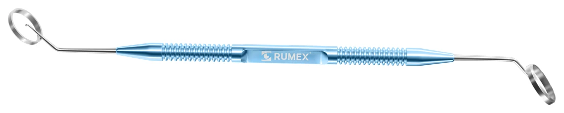
Corneal marker
3-0231
-

Mendez degree gauge
2-0331T
-

Tunnel zone marker
3-143T
-

Diamond knife
6-00 / 6-020
-

ICRS elevator
10-035
-
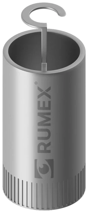
4.5 mm CAIRS dissector
16-175
-
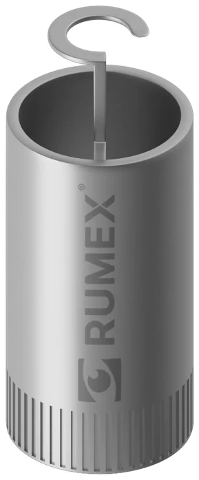
5.5 mm CAIRS dissector
16-176
-
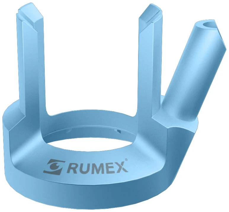
Vacuum stabilisation guide
16-315T
-

Suarez spreader
13-146
-

Bicalto guide
13-147
-

Castroviejo fixation forceps
4-0822T
-

McPherson tying forceps
4-178S
-

Reversed Sinskey hook
5-0322
-

Right Y pusher
5-080
-

Left Y pusher
5-081
-

Thornton fixation ring
16-036S
-
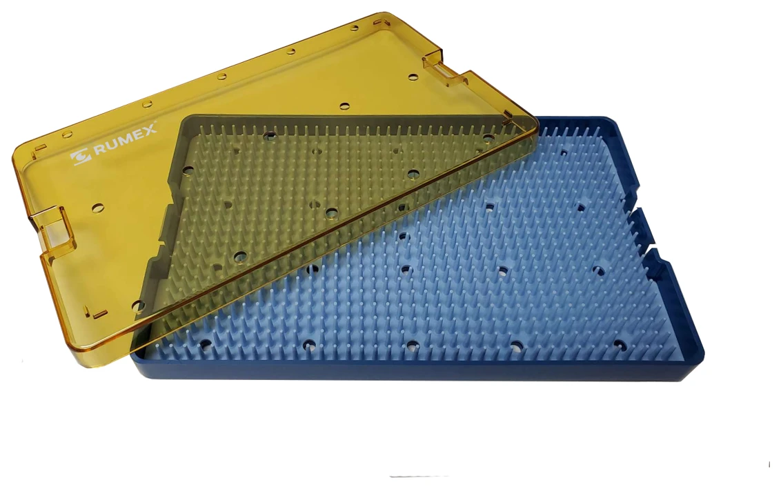
Sterilisation tray
18-304
Instrument photographs: SRB Devices, Madhu Instruments, Epsilon USA, Rumex, Dr Soosan Jacob and Dr Aylin Kılıç. CLEAR marker in use: Ziemer technique supplement.
Complete the theatre set
Alongside the dedicated CAIRS tools, prepare the routine instruments and consumables for your chosen method: corneal marking and angle measurement, fine tissue forceps, scissors for arc-length trimming, and the tissue preparation surface or artificial anterior chamber where required. Manual channel creation needs its own trained dissection workflow and instruments.
Practise with the exact instruments you intend to use before your first patient. Familiarity with their curvature, direction and tissue grip makes it easier to recognise what is happening during insertion.
Teaching and further reading
Instrument roles follow the CAIRS teaching library. Product details are drawn from the supplier and designer sources linked below; confirm current sizes and ordering details with the supplier.
- SRB Devices — Cronin–Gunn CAIRS instruments
- Madhu Instruments — Jacob CAIRS trephine
- Epsilon USA — Awwad CAIRS set
- Soosan Jacob — customised segment preparation video
- Shady Awwad — CAIRS micrograspers video
- Awwad and colleagues — published preparation and implantation technique
- KeraNatural — AK instrument list and distributor details (PDF)
- Aylin Kılıç — AK forceps, tunnel separators and marker
- Rumex — LWVI CAIRS treatment set for manual surgery